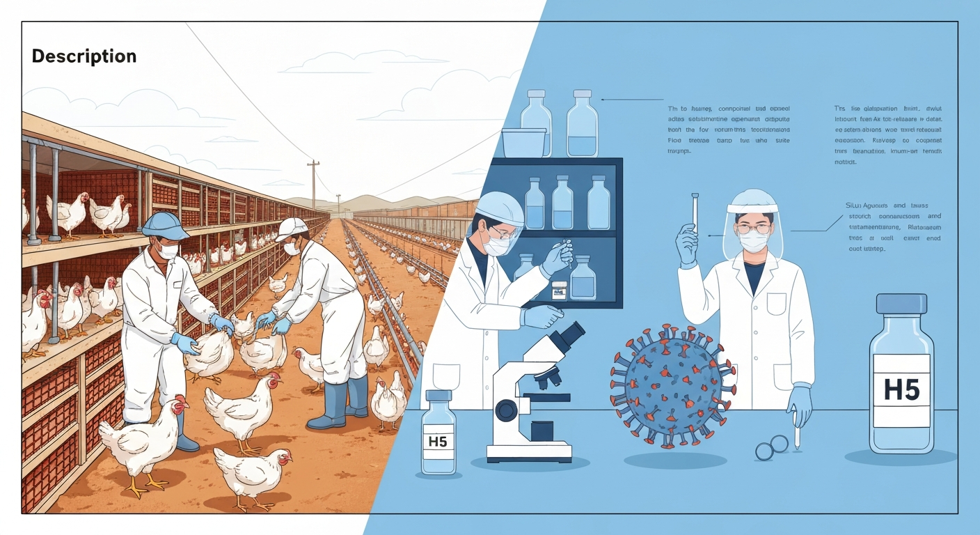
Bird flu is caused by an RNA virus, not a DNA virus. Design and Performance of the CDC Real‑Time RT‑PCR Swine Flu Panel, CDC / Journal of Clinical Microbiology notes that CDC/FDA influenza real‑time RT‑PCR diagnostic panels are designed to detect influenza A (universal assays commonly target the conserved matrix (M) gene) and include primer/probe sets for subtyping (HA/NA or specific subtypes); these assays are validated for respiratory specimen types Design and Performance of the CDC Real‑Time RT‑PCR Swine Flu Panel — CDC / Journal of Clinical Microbiology. Specifically, avian influenza viruses belong to the family Orthomyxoviridae and carry a single-stranded, negative-sense RNA genome split across eight separate segments. That combination of RNA-based replication and a segmented genome is the root cause of nearly everything that makes influenza unpredictable: its high mutation rate, its ability to generate entirely new strains through reassortment, and the reason why diagnostic labs need a special reverse transcription step before they can run a standard PCR test.
Is Bird Flu RNA or DNA? Understanding Avian Influenza

What is avian influenza (bird flu)?
Avian influenza is an infectious viral disease caused by influenza A viruses that circulate naturally in wild aquatic birds, particularly waterfowl such as ducks, geese, and shorebirds. These birds often carry the virus without showing symptoms, which allows it to spread silently across flyways and between continents. When the virus jumps into domestic poultry such as chickens and turkeys, the consequences can be severe: highly pathogenic avian influenza (HPAI) strains, notably H5N1 and the currently circulating H5N1 clade 2.3.4.4b, cause catastrophic mortality in flocks. The virus also infects mammals, including cats, mink, sea lions, and cattle, and occasional human infections do occur, typically in people with direct, prolonged exposure to infected birds or contaminated environments. Sustained human-to-human transmission has not been established, which is why HPAI has not yet caused a human pandemic, but the virus's genetic makeup means that risk must be monitored continuously.
The structure and genome of influenza: eight RNA segments
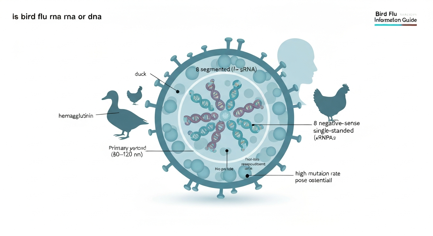
An influenza A virus particle is roughly spherical, about 80 to 120 nanometers in diameter, and wrapped in a lipid envelope studded with two key surface proteins: hemagglutinin (HA) and neuraminidase (NA). It is those two proteins that give each influenza A strain its name (H5N1, H7N9, and so on). There are currently 18 known HA subtypes and 11 known NA subtypes in nature.
Inside the envelope, the genetic material is not packaged as a single continuous strand. Instead, the influenza A genome consists of eight distinct RNA segments, each encoding one or more viral proteins and each wrapped in viral proteins to form ribonucleoprotein complexes (vRNPs). The eight segments code for: the three RNA polymerase subunits (PB2, PB1, and PA), hemagglutinin (HA), nucleoprotein (NP), neuraminidase (NA), the matrix proteins (M1 and M2), and the non-structural proteins (NS1 and NEP). Each segment is a linear, single-stranded, negative-sense RNA molecule, which means it is the mirror image of the messenger RNA the cell would read. The virus must first convert it into a readable form before it can make new proteins.
| Genome Segment | Protein(s) Encoded | Key Function |
|---|---|---|
| 1 (PB2) | PB2 polymerase subunit | Cap binding during RNA synthesis |
| 2 (PB1) | PB1 polymerase subunit | RNA-dependent RNA polymerase catalytic core |
| 3 (PA) | PA polymerase subunit | Endonuclease activity; RNA synthesis |
| 4 (HA) | Hemagglutinin | Cell attachment; vaccine target |
| 5 (NP) | Nucleoprotein | Encapsidates RNA; nuclear transport |
| 6 (NA) | Neuraminidase | Releases new virions from host cell |
| 7 (M) | M1, M2 | Structural matrix; ion channel |
| 8 (NS) | NS1, NEP | Interferon antagonist; nuclear export |
Why RNA vs. DNA matters: mutation, drift, and reassortment
The distinction between RNA and DNA is not just academic. It has direct consequences for how quickly bird flu evolves and how difficult it is to control.
High mutation rate and antigenic drift
The enzyme influenza uses to copy its RNA genome is called RNA-dependent RNA polymerase (RdRp). Unlike the DNA polymerases that replicate human chromosomes, RdRp lacks a proofreading mechanism. It cannot detect and fix copying errors in real time. The result is a mutation rate roughly in the range of 10 to the minus 6 through 10 to the minus 4 substitutions per nucleotide per replication cycle, which translates to evolutionary rates for key surface proteins like HA on the order of 5 to 7 thousandths of a substitution per site per year. That sounds small, but across billions of viral replication cycles, it produces a constant stream of variants. The gradual accumulation of these small changes in the HA and NA proteins is called antigenic drift. It is the reason seasonal flu vaccines need to be updated regularly and the reason surveillance labs around the world sequence influenza viruses year-round through WHO's Global Influenza Surveillance and Response System (GISRS).
Reassortment and antigenic shift
The eight-segment genome creates a second, more dramatic source of change. When two different influenza A strains infect the same cell at the same time, the packaging machinery can grab segments from either virus. The resulting progeny may carry a completely new combination of segments, including an HA or NA subtype that circulating human immune systems have never encountered. This process is called reassortment, and when it produces a virus with dramatically different surface proteins, it is the mechanism behind antigenic shift. Pandemic strains have repeatedly emerged this way: the 1957 H2N2 pandemic strain acquired three new gene segments from an avian source, and the 2009 H1N1 pandemic virus was a reassortant of human, swine, and avian lineages. Pigs are considered mixing vessels for reassortment because their respiratory tracts carry receptors for both avian and human influenza viruses, but any animal co-infected with two strains can theoretically generate new combinations.
How genetic features shape transmission risk and virus evolution
Antigenic drift and reassortment do not just matter theoretically. They are the reason that H5N1 has been monitored so intensely since its re-emergence in 2021 under clade 2.3.4.4b. That particular lineage has spread to an unprecedented number of mammalian species, including cattle in the United States, raising concern that it is finding footholds in new hosts. Each new host species is an opportunity for the virus to accumulate mutations that improve replication in mammalian cells or, more worryingly, to co-infect with a human influenza strain and undergo reassortment. Mutations in PB2, a polymerase subunit encoded by segment 1, are particularly watched because specific substitutions at positions like 627K and 701N are associated with enhanced replication at the lower temperatures of the mammalian respiratory tract. When surveillance labs detect these markers, it triggers escalated response from public health agencies.
For the general public and for poultry workers, the practical implication is that transmission risk is not static. A strain that poorly infects humans today might acquire a handful of mutations that change that picture. That is why regular monitoring, rapid reporting of sick or dead birds, and occupational precautions for farm workers are treated as essential public health infrastructure rather than optional measures.
How bird flu is detected and diagnosed
Because the influenza genome is RNA, detecting it requires a different approach than standard DNA-based PCR. Here is how the main methods work in practice.
Real-time RT-PCR: the front-line molecular test
The workhorse diagnostic is real-time reverse transcription PCR (rRT-PCR or RT-PCR). The test works in two stages. First, an enzyme called reverse transcriptase converts the viral RNA into complementary DNA (cDNA), a form that standard PCR machinery can amplify. Then PCR amplifies the cDNA exponentially while a fluorescent probe signals when target sequence is present. The CDC's validated influenza RT-PCR panels typically target the conserved matrix (M) gene for universal influenza A detection, with additional primer-probe sets to subtype the HA and NA proteins. This approach achieves sensitivity well above 90% in properly collected and handled specimens, making it the recommended test for hospitalized patients and anyone with potential H5N1 exposure.
Specimen types and why timing matters
For humans, the recommended specimens are upper respiratory swabs: nasopharyngeal, nasal, or throat swabs collected as early in illness as possible, ideally within the first 3 to 4 days of symptom onset when viral loads are highest. In severe or hospitalized cases, lower respiratory specimens such as bronchoalveolar lavage (BAL) fluid increase detection sensitivity. For birds, oropharyngeal and cloacal swabs are collected simultaneously because avian influenza viruses replicate in both the respiratory and gastrointestinal tracts of birds. RNA degrades quickly at room temperature, so swabs must be placed in viral transport medium, kept cold, and processed promptly. A negative RT-PCR result from a specimen collected too late or handled incorrectly does not rule out infection.
Rapid antigen tests: useful but limited
Rapid influenza diagnostic tests (RIDTs) detect viral antigen rather than RNA and can produce a result in 10 to 15 minutes. They are useful for quick triage in clinical settings, but their sensitivity is substantially lower than RT-PCR: pooled estimates from meta-analyses place RIDT sensitivity at roughly 50 to 70% compared with molecular reference standards, with higher specificity. The practical implication is that a negative rapid antigen test in a high-risk patient should not be treated as definitive. CDC guidance specifically recommends confirming negative RIDT results with RT-PCR in hospitalized patients or anyone with suspected avian influenza exposure.
Whole-genome sequencing and serology
Once a sample tests positive by RT-PCR, public health laboratories often proceed to whole-genome sequencing (WGS). Sequencing all eight gene segments simultaneously provides detailed information about the strain's subtype, phylogenetic lineage, and any mutations of concern, including those associated with antiviral resistance or mammalian adaptation. This data feeds directly into WHO's GISRS network and informs decisions about vaccine strain composition. Serological tests (measuring antibodies against influenza in blood samples) are used epidemiologically, particularly to understand how widely a strain has circulated in a population, but they are not useful for acute clinical diagnosis because antibody levels take one to two weeks to rise after infection. Virus isolation in embryonated eggs or MDCK cell lines remains the classical gold standard and is still used for confirmatory work and vaccine strain production, but it requires higher biosafety containment for HPAI and is far slower than molecular methods.
| Test Type | Target | Sensitivity vs. RT-PCR | Turnaround | Best Use |
|---|---|---|---|---|
| Real-time RT-PCR | Viral RNA | Reference standard (>90%) | 2–6 hours | Clinical diagnosis, outbreak confirmation |
| Rapid antigen test (RIDT) | Viral protein antigen | ~50–70% | 10–15 minutes | Quick triage; confirm negatives in high-risk cases |
| Whole-genome sequencing | All 8 RNA segments | N/A (characterization) | Days to weeks | Surveillance, strain characterization, vaccine selection |
| Serology (antibody test) | Host antibodies to virus | N/A (retrospective) | Days | Seroprevalence studies, exposure investigations |
| Virus isolation (culture) | Live virus | High (slower) | Days to weeks | Confirmatory, vaccine seed stock production |
Why RNA viruses need a different diagnostic approach
The reverse transcription step in RT-PCR is not just a technical detail. It reflects something fundamental about RNA virology. Standard PCR requires a DNA template. Because influenza carries RNA, you cannot skip straight to amplification. The RT step is also where many diagnostic failures happen: reverse transcriptase is sensitive to inhibitors in biological specimens, and RNA degrades more readily than DNA when samples are mishandled. This is why laboratory guidance stresses cold-chain management and recommends processing respiratory specimens within 24 to 72 hours of collection, or storing them frozen at minus 70 degrees Celsius if longer delays are unavoidable. For anyone coordinating specimen collection in the field, whether on a poultry farm or in a rural clinic, understanding that you are working with an unstable RNA target is important context for following those handling protocols carefully.
Bird flu vaccines: types, development timelines, and who makes them
Vaccine development for avian influenza is closely tied to the same RNA biology discussed above, because the vaccines must target the HA protein, which keeps drifting. Here is a practical overview of where things stand. See when was the bird flu vaccine made for a brief timeline of key dates in the vaccine's development.
Current vaccine types and platforms
Human pandemic preparedness vaccines for H5N1 and other avian influenza subtypes have been developed using several platforms. Traditional inactivated vaccines are grown in embryonated chicken eggs, blank" rel="noopener noreferrer">a process that typically takes around six months from strain selection to distributed doses. Cell-based vaccines use mammalian cell lines instead of eggs and can shorten some manufacturing steps. Recombinant protein vaccines express the HA antigen in insect or other cell systems without any live virus, offering another alternative. More recently, mRNA vaccine platforms, the same technology used in COVID-19 vaccines, have been applied to influenza. mRNA vaccine candidates can be designed within days of identifying a target sequence, though manufacturing scale-up and regulatory review still add considerable time before doses are ready for widespread use. If you want to know how long did bird flu vaccine take to develop, initial vaccine designs can be made within days but full clinical testing, regulatory review, and manufacturing scale-up typically take months to a year depending on the platform and urgency. Questions about whether a bird flu vaccine would use mRNA technology are reasonable given the attention that platform received during the pandemic, and several H5N1 mRNA vaccine candidates have entered or are approaching clinical trials.
Who makes bird flu vaccines and WHO's role
Multiple pharmaceutical manufacturers hold or are developing pre-pandemic H5N1 vaccine stockpiles, including Sanofi, CSL Seqirus, and others working under contracts with national governments. See who makes bird flu vaccine for more details on manufacturers and stockpiles. The U.S. government maintains a Strategic National Stockpile of H5N1 vaccine doses. WHO plays a coordinating role through GISRS, meeting twice yearly to recommend which strains should be used for vaccine composition for both seasonal and pandemic preparedness purposes. WHO and bird flu vaccine guidance through GISRS informs strain selection and pandemic preparedness. National regulatory authorities make the final approval decisions, but WHO's recommendations are the backbone of the global process. For humans, avian influenza vaccines are not currently recommended for the general public. For information on how to get bird flu vaccine, including who is eligible and where to obtain doses, consult official public health guidance. Vaccination guidance focuses on occupationally exposed individuals such as poultry workers, farm veterinarians, and laboratory personnel working with HPAI viruses.
How effective are bird flu vaccines?
Effectiveness data for avian influenza vaccines in humans is more limited than for seasonal flu because widespread use has not occurred. Pre-clinical and clinical immunogenicity trials show that candidate H5N1 vaccines can generate protective antibody responses, particularly when given with an adjuvant or as a two-dose series. Seasonal influenza vaccine effectiveness, by comparison, typically ranges from about 40 to 60% against matched strains, and lower when strains drift from vaccine composition. The honest answer on pandemic bird flu vaccine effectiveness is that the true figure will depend heavily on how well the circulating pandemic strain matches the vaccine antigen, which is why global sequence surveillance through GISRS is so important. For more detail on vaccine performance in trials and likely effectiveness in pandemic scenarios, see how effective is bird flu vaccine.
Public health implications: transmission routes, food safety, and human risk
Most people who have contracted H5N1 had direct, close contact with infected live poultry or heavily contaminated environments. Casual contact with birds does not carry the same level of risk, and properly cooked poultry and eggs are safe to eat: influenza viruses are inactivated at the internal cooking temperatures recommended for poultry (74 degrees Celsius or 165 degrees Fahrenheit). You should not handle dead or visibly sick wild birds with bare hands, and if you find multiple dead birds in a localized area, reporting them to your state or local wildlife or agriculture agency is the appropriate step.
Farm workers and veterinarians who are regularly in contact with poultry or potentially infected animals represent the highest-risk group for human exposure. The CDC recommends that these individuals use appropriate personal protective equipment including respirators (N95 or higher), eye protection, gloves, and protective clothing when working with or near infected or potentially infected animals. They should also be current on seasonal influenza vaccination, partly for their own protection and partly to reduce the theoretical possibility of co-infection with a human influenza strain that could enable reassortment.
When to seek care and how to report concerns
Anyone who has had direct contact with infected or suspected poultry and develops fever, respiratory symptoms, or conjunctivitis within 10 days of exposure should contact a healthcare provider promptly and mention the potential exposure history. Antiviral medications like oseltamivir (Tamiflu) are most effective when started early in the course of illness, so prompt medical evaluation matters. Do not wait for a rapid antigen test result to rule out avian influenza in that scenario.
- Report dead or sick wild birds (especially waterfowl or raptors) to your state wildlife agency or USDA APHIS.
- Report sick or dying poultry to your state veterinarian or local animal health official immediately.
- If you have had poultry exposure and develop fever or respiratory symptoms within 10 days, call a healthcare provider and describe the exposure before visiting a clinic.
- Poultry workers should use N95 respirators, eye protection, gloves, and protective outerwear when working in affected flocks or during depopulation.
- Stay current on seasonal influenza vaccination if you work with animals or in a healthcare setting.
- Do not touch dead wild birds with bare hands; use gloves or a bag inverted over your hand.
The RNA nature of avian influenza is not just a trivia answer. It explains the virus's capacity to change, the way labs detect it, and the challenges vaccine developers face in staying ahead of it. Understanding those connections helps make sense of why public health agencies invest so heavily in surveillance, rapid diagnostics, and pandemic preparedness even when human cases remain rare. The biology is the foundation of the response.
FAQ
Is bird flu RNA or DNA?
Bird flu (avian influenza) viruses are RNA viruses. Specifically, influenza A viruses—those most often referred to as “bird flu”—have single‑stranded, negative‑sense RNA genomes that are segmented into eight separate RNA segments (not DNA).
What does negative‑sense, single‑stranded RNA mean?
Negative‑sense RNA means the viral genome is the complement of the messenger RNA (mRNA) needed to make viral proteins. The virus carries an RNA‑dependent RNA polymerase that first makes positive‑sense copies (mRNA) from the negative‑sense genome so proteins can be produced and new viruses assembled.
Why does it matter that bird flu is an RNA virus?
RNA genomes generally mutate faster than DNA because their polymerases lack strong proofreading. This higher mutation rate drives antigenic drift (gradual change). The segmented genome also allows reassortment—when two different influenza viruses infect the same cell they can swap whole genome segments—producing antigenic shift (sudden emergence of novel strains). Both processes affect transmissibility, host range, and vaccine match.
How do these RNA properties affect transmission risk to humans?
Higher mutation rates and reassortment increase the chance that an avian virus could acquire changes that help it infect humans or transmit more easily. Most avian influenza viruses do not transmit efficiently between people, but monitoring for genetic changes helps public health detect strains with increased human risk early.
How is bird flu detected and diagnosed?
Laboratory diagnosis is typically by nucleic acid amplification tests that start with reverse transcription (RT) to convert viral RNA into DNA, followed by PCR (real‑time RT‑PCR or rRT‑PCR). These tests are sensitive and specific. Rapid antigen tests exist but have lower sensitivity and negative results in high‑risk cases should be confirmed by molecular testing. Virus isolation in eggs or cell culture and whole‑genome sequencing are used for surveillance and characterization.
Why do tests for bird flu use RT‑PCR instead of regular PCR?
Because the virus genome is RNA, a reverse transcription step is required to make complementary DNA before PCR amplification. Regular PCR amplifies DNA, so RT‑PCR (or rRT‑PCR) is the standard molecular method for RNA viruses like influenza.
When Was the Bird Flu Vaccine Made? Timeline & Facts for Birds and People
Learn when bird‑flu vaccines first appeared for poultry and humans, how they work, effectiveness, and who can get them.